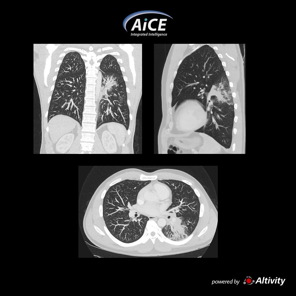

Pneumonia seen in these MPR images of this 49-year-old man with shortness of breath and BMI 25.4
Courtesy
South Coast Radiology
Qld, Australia
View Scan Parameters
Scan ModeUltra Helical
Collimation0.5 mm × 80
kVp120
mAsSUREExposure
Rotation Time0.35 s
Scan Range409 mm
Dose ReductionAiCE
CTDIvol4.5 mGy
DLP204.4 mGy·cm
Effective Dose2.9 mSv
k-factor0.014 (AAPM Report 96)
×
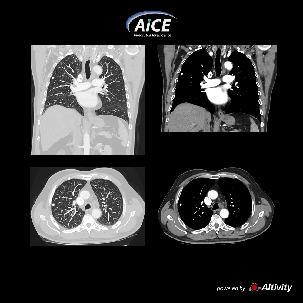
Chest – Calcified Nodule
82-year-old man, follow up chest CT demonstrates a calcified lung nodule in the right lung
Courtesy
South Coast Radiology
Qld, Australia
View Scan Parameters
Scan ModeUltra Helical
Collimation0.5 mm × 80
kVp120
mAsSUREExposure
Rotation Time0.35 s
Scan Range364.5 mm
Dose ReductionAiCE
CTDIvol5.2 mGy
DLP214 mGy·cm
Effective Dose3.0 mSv
k-factor0.014 (AAPM Report 96)
×
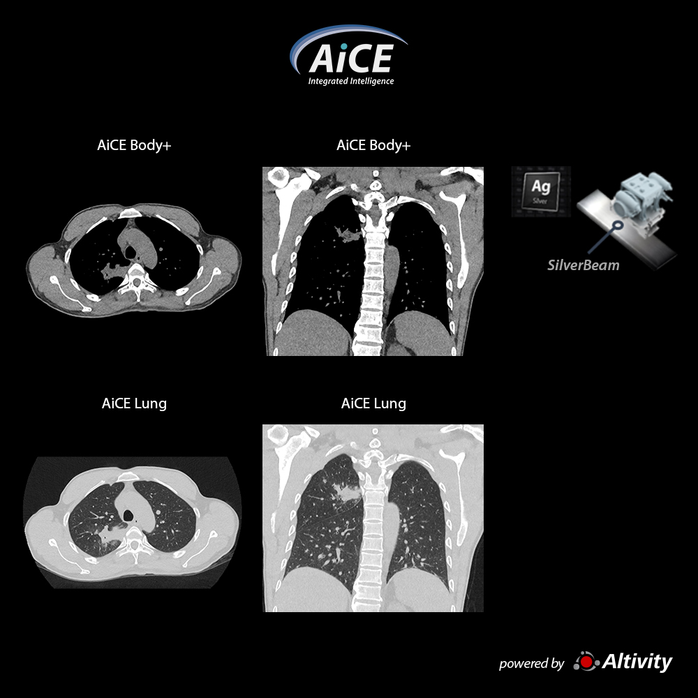
Low dose chest with SilverBeam
SilverBeam in combination with AiCE Deep Learning Reconstruction provides excellent image quality at dose levels more comparable to a chest x-ray.
In this patient the DLP was only 24 mGy.cm with an effective dose of 0.3 mSv.
Courtesy
Medic Medical Center, HCMC Vietnam
View Scan Parameters
Scan ModeSilverBeam Ultra-Helical
Collimation0.5 mm × 80
kVp120
mAsSUREExposure
Rotation Time0.5 s
Scan Range241 mm
Dose ReductionAiCE
CTDIvol0.70 mGy
DLP24.70 mGy-cm
Effective Dose0.34 mSv
k-factor0.014 (AAPM Report 96)
×
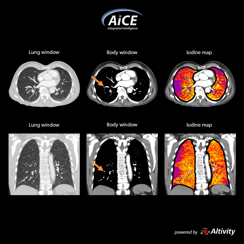
Pulmonary CTA - Iodine Mapping SCT
Color iodine maps quickly and definitively show areas of hypoperfusion to enhance your diagnosis.
Hyperperfused areas are commonly associated with pulmonary pathology as demonstrated in this case of a pulmonary embolism.
Courtesy
Royal Bournemouth Hospital, Bournemouth, UK